Our Services

As a fully licensed and equipped optometric practice, Dr. Mungar offers a complete range of eye care services to all our patients.
Whether the eye care issue involves correcting refractive errors with eyeglasses or contact lenses or helping a student find frames that will not look geeky, our experienced team will identify and implement the best eye care solutions possible.
Eye Health Evaluation
With our years of experience in diagnosing and treating typical vision disorders such as nearsightedness, farsightedness, amblyopia, presbyopia, cataracts, macular degeneration, and diabetic retinopathy, Dr. Mungar is also licensed to provide appropriate therapeutic medical eye care.
At the same time, our practice offers a wide array of the very best high-quality eye care products at reasonable prices. Our patients never pay too much for the best quality eyeglasses, contact lenses, sunglasses, progressive and bifocal lenses, and outstanding service.
Treatment of Eye Disease
If you are diagnosed with an eye disease, you want only the best treatment available to get your eyes healthy again. Dr. Mungar offers only the best. Based on your diagnosis, we may recommend a wide variety of approaches, including improved nutrition, prescription medicines, therapy and vision exercises, or medical procedures.
Good Eyecare Begins With A Yearly Eye Exam!
Although many do not realize it, the best way to protect your vision is with a yearly eye checkup. Even a basic eye exam can instantly detect such health-threatening conditions as tumors, vascular irregularities, and diabetes-related injury to the retina. With Dr. Mungar, that basic checkup is comprehensive and takes less than a ½ hour.
Digital Retinal Imaging
In our continued efforts to bring the most advanced technology available to our patients, Dr. Mungar is proud to announce the inclusion of the Digital Retinal Imaging as an integral part of your eye exam.
Many eye problems can develop without warning and progress with no symptoms. Early on, you might not notice any change in your vision. However, diseases such as macular degeneration, glaucoma, retinal tears or detachments, as well as other health problems such as diabetes and high blood pressure, can be detected with a thorough exam of the retina. The retina is the part of your eye that catches the image of what you are looking at, similar to the film in a camera.
Digital Retinal Imaging provides:
- A digital image to confirm a healthy eye or detect the presence of disease.
- A more wide-field view can be achieved than a regular examination can provide on it’s own.
- The opportunity for you to view and discuss the images of your eye with your doctor at the time of your exam.
- A permanent record for your medical file, enabling your optometrist to make important comparisons if potential problems show themselves at a future examination.
LASIK
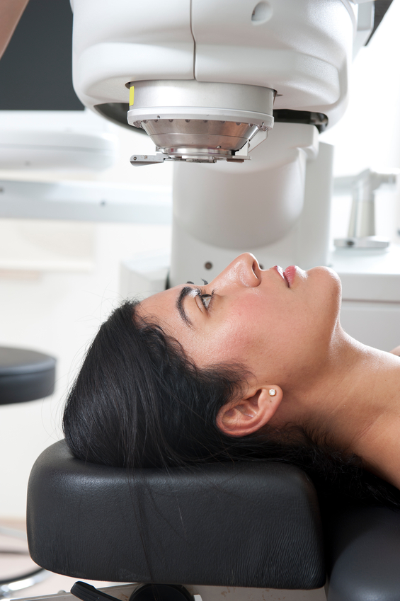
LASIK, a form of refractive surgery, is an popular option for vision correction, often eliminating the need to wear glasses or contact lenses. Simply put, LASIK reshapes the cornea with a laser.
Other surgical alternatives have become available. Among these is a technique called phakic IOL implantation which involves implanting a lens behind the cornea, but in front of the iris. With this new option, many of those who were too highly nearsighted for LASIK are now candidates for refractive surgery.
If you are interested in refractive surgery, please let us know. Refractive surgery is not to be taken lightly. Detailed testing is necessary to determine whether or not you are a good candidate for the surgery. If testing shows you to be a good candidate, we can help you choose the refractive surgeon who is most appropriate for your case. In addition, we provide post-operative care for refractive surgery.
BlephEx Eyelid Microblepharoexfoliation
We are excited to offer BlephEx treatment to our patients in the battle against blepharitis and dry eye. Blepharitis and or dry eye affect upwards of half of the adult population. The BlephEx procedure removes the biofilm from the lid margins and lashes, decreasing the quantity of bacteria in these areas. The microexfoliation also exposes the tiny openings of the meibomian glands, allowing for better lipid release onto the tear layer.
Please use the following links to learn more:
LipiFlow
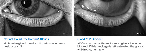
LipiFlow is an FDA approved medical device for treating meibomian gland disease.
Meibomian gland disease or dysfunction (MGD) refers to the condition where the meibomian glands are not secreting enough oil or when the oil they secrete is of poor quality. Often, the oil gland openings get blocked so that less oil comes out of the gland. The oil that does make it out of the glands can be thickened and unhealthy and can cause irritation. It is believed that 86% of dry eye cases are due to MGD.
The LipiFlow System represents more than 10 years of dedicated research and is protected by more than 30 patents. It uses precisely controlled therapeutic heat and massage to relieve gland blockages. LipiFlow is safe and pain-free.
Patented components called Activators deliver treatments directly to the inner and outer eyelids while protecting delicate eye tissue. Sterile and single-use, the Activators are designed for safety and comfort.
Each treatment takes about 12 minutes. Most patients find improvement of their symptoms in as little as two weeks, with the results lasting 12 months or more. Dr. Mungar and his staff can guide you on what to expect.
Optical Coherence Tomography / OCT
When patients come to our office for eye exams, many times there is testing that we do to help us diagnose problems. One of these tests uses Optical Coherence Tomography or OCT. Pictures taken with our OCT machine are generated by light waves that reflect off the back of the eye or retina, creating images similar to what could be produced by a low-power microscope. The OCT also provides cross-sectional images. These images can display the various layers of the retina. This technology is also used to image the optic nerve, which is important in glaucoma treatment and management.
OCT is a non-invasive and no-contact test that doesn't require preparation from the patient. There is no exposure to radiation since the machine uses light to obtain the images. The patient sits in front of a machine, a couple of bright flashes like a normal camera flash go off, and then the photos can be viewed on the machine within a minute.
The OCT is an extremely valuable tool used to help diagnose and manage common retinal eye diseases such as macular degeneration, macular edema (fluid in the retina), and macular hole/epiretinal membranes. We use the initial OCT images to aid in making a definitive diagnosis. The OCT compares these initial images of your eye to a database of images of normal eyes matched to your age. In this way, the first OCT images we take can help point out potential issues if there is something that looks different from normal or average. Subsequent OCT images can then compare how you look now compared to how you looked initially. This can be very valuable in gauging how well treatment is working or if the problem is progressing.
The other common use of the OCT is for monitoring and managing glaucoma. We usually take initial images of the optic nerve, and the OCT can then compare these images to those of age-matched healthy control patients. The OCT will usually be repeated every year so we can follow any changes or progression over time.
The advent of OCT has revolutionized the way we evaluate the retina because we can now detect subtle findings not otherwise easily seen during clinical exams. This makes the OCT one of the most valuable tests we can do in our office.